ImageJ - Free Image Processing and Analysis Software

ImageJ is one of the most widely used and powerful free software tools for viewing, processing, and analyzing digital images. It is particularly popular in scientific research, medicine, engineering, and other technical fields. Originally developed at the National Institutes of Health (NIH), ImageJ provides a comprehensive collection of image processing and analysis tools in a lightweight and user-friendly environment. It allows users to perform a wide range of image analysis tasks without requiring any programming knowledge.
One of the key advantages of ImageJ is its lightweight design and ease of use. Unlike many specialized image processing applications that require substantial hardware resources, ImageJ has a relatively small footprint and low memory requirements, allowing it to run on a wide range of computer systems. Portable versions are also available, making it possible to run the software directly without a conventional installation in many cases. These features make ImageJ a practical choice for students, researchers, engineers, and professionals who need a fast and accessible tool for image inspection and analysis.
Image Viewing and Image Management
ImageJ supports loading, viewing, and managing images in a variety of file formats. It is compatible with common formats such as JPEG, PNG, TIFF, and BMP, as well as specialized scientific image formats. The software also supports images with different bit depths and multiple channels, allowing users to examine image data in greater detail.
In addition to conventional images, ImageJ can open and process multi-page images and image stacks. This feature is particularly useful for analyzing microscopy images, medical scans, and image sequences acquired at different time points or spatial positions. For example, a series of consecutive images can be loaded as a single stack, allowing users to navigate between individual slices or apply processing operations to the entire image sequence.
Image Processing and Editing Tools
ImageJ offers an extensive collection of tools for image processing and editing. Users can perform common operations such as cropping, resizing, rotating images by arbitrary angles, repositioning, and adjusting image dimensions. The software also supports changing image data types and converting between 8-bit, 16-bit, and 32-bit formats.
Other important features include brightness and contrast adjustment, modification of pixel intensity ranges, application of various image filters, and noise reduction. These tools help users improve image appearance and enhance specific details for more accurate visual inspection and subsequent analysis.
ImageJ also provides a range of spatial and frequency-domain processing tools. Filters such as Mean, Median, and Gaussian Blur, along with other processing operations, can be used to reduce noise, smooth images, or enhance specific structures and features.
Image Segmentation and Thresholding
One of ImageJ’s useful capabilities is its collection of selection and segmentation tools. Users can manually define regions of interest (ROIs) using different selection tools or employ automated and semi-automated methods to distinguish specific structures from their surroundings.
The Thresholding tool allows users to define pixel intensity ranges and separate regions of interest from the background. This functionality is widely used in microscopy, laboratory image analysis, and medical image processing. Once thresholding is applied, the selected regions can be displayed as binary masks and used for further measurements and quantitative analysis.
Image Measurement and Quantitative Analysis
ImageJ is more than just an image viewer and editor; it also provides powerful tools for extracting quantitative information from digital images. Users can measure parameters such as area, perimeter, length, angle, mean pixel intensity, minimum and maximum intensity, and various other statistical properties.
An important feature of ImageJ is its spatial calibration capability. When the physical scale of an image is known, pixel-based measurements can be converted into real-world units such as millimeters, micrometers, or other measurement units. This is particularly important for accurate measurements in microscopy and medical imaging applications.
Users can also draw lines, circles, rectangles, and other geometric shapes to define specific regions and measure their properties. Measurement results are displayed in a dedicated results table and can be exported to text files or CSV format for further analysis.
Plugins and Extensibility
One of the main reasons behind ImageJ’s widespread adoption is its extensible architecture. The software supports plugins, allowing users to expand its functionality by installing additional tools. A large collection of plugins has been developed for specialized image processing tasks, including advanced segmentation, three-dimensional image analysis, image reconstruction, and specialized measurement techniques.
ImageJ also supports macros, which allow users to automate repetitive operations and apply standardized processing workflows to large numbers of images. This feature is particularly valuable for researchers working with large datasets who need to perform consistent analyses across multiple images.
For more advanced users, ImageJ also provides the ability to develop custom tools and plugins using the Java programming language. Therefore, although no programming knowledge is required for routine image processing and analysis, ImageJ can also serve as a highly customizable development environment for specialized imaging applications.
Applications of ImageJ in Medical Imaging and Industry
ImageJ is used across a wide range of scientific and technical disciplines. In biological and microscopy research, it is commonly used for measuring cellular structures, examining tissue samples, counting particles, and analyzing fluorescence images. In medicine and medical imaging, it can be used to view and process images acquired using X-ray and C-arm imaging, computed tomography (CT), magnetic resonance imaging (MRI), and other digital imaging modalities.
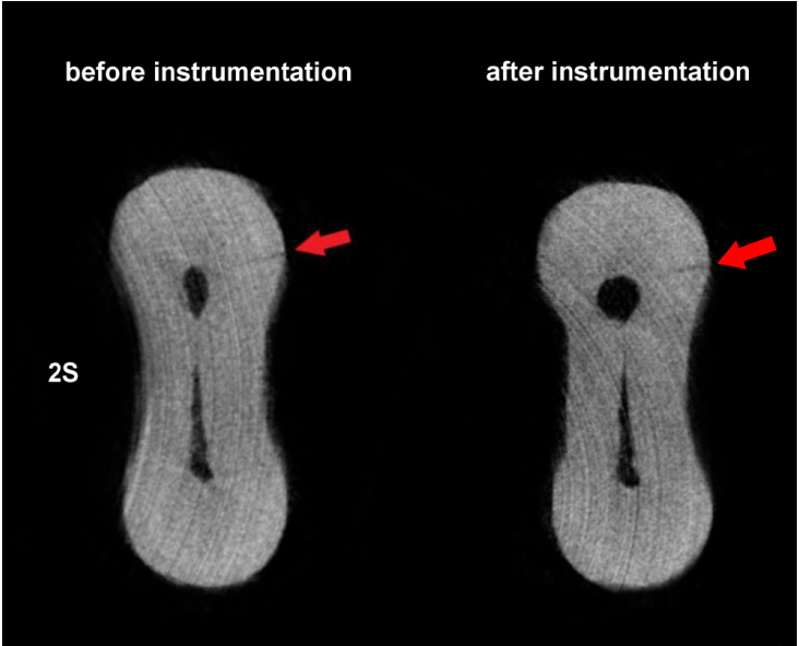
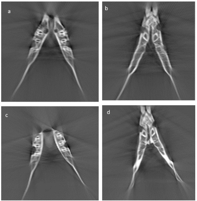
One particularly important application of ImageJ is the analysis of micro-computed tomography (Micro-CT) images. Micro-CT is an X-ray-based imaging technique that enables high-resolution, three-dimensional visualization of the internal structures of objects and specimens. It is widely used in preclinical research, dental research, tissue engineering, pharmaceutical and pharmacological studies, and the investigation of microscopic structures.
ImageJ provides a range of tools for processing and analyzing Micro-CT datasets and extracting valuable quantitative information. These include thresholding and segmentation of bone tissue from the background, isolation of specific anatomical structures, measurement of bone area and volume, assessment of relative density, and analysis of morphometric parameters such as trabecular thickness (Tb.Th), trabecular separation (Tb.Sp), and bone volume fraction (BV/TV).
With specialized plugins such as BoneJ, users can also perform more advanced analyses of three-dimensional bone architecture and trabecular microstructure.
The Advanced X-ray Imaging Systems (AXIS) Laboratory at Behin Negareh is the country’s first specialized laboratory dedicated to providing high-quality Micro-CT imaging and quantitative and qualitative analysis services for researchers across various scientific disciplines. Examples of published research using Micro-CT systems manufactured by Behin Negareh, including LOTUS-inVivo and LOTUS-NDT, with image analysis performed by the laboratory’s specialists, are available in the published papers section. Information about available imaging and analysis services can also be found in the service and price list.
Beyond Micro-CT, ImageJ can be used in many other imaging applications. For example, researchers can examine signal intensity across different image regions, measure anatomical structures, isolate specific areas, and extract statistical features from selected regions of interest. Its ability to handle multi-slice image stacks also makes it a useful tool for analyzing volumetric image datasets.
It is worth noting that ImageJ is not intended to replace specialized diagnostic software or clinically validated medical imaging systems. However, it offers a valuable set of tools for research, education, and quantitative image analysis.
Why ImageJ?
ImageJ combines several important advantages, including its free and open-source availability, lightweight design, ease of use, support for a wide range of image formats, and extensive collection of image processing and analysis tools. Most routine research and educational tasks can be performed without requiring high-end computer hardware. Furthermore, its extensive plugin ecosystem allows users to expand its capabilities to meet the requirements of different scientific and technical applications.
Overall, if you are looking for a lightweight, free, and powerful solution for viewing, editing, processing, and quantitatively analyzing digital images, ImageJ is an excellent choice. Downloading ImageJ gives you access to a versatile image analysis environment that supports a wide range of processing operations, with the flexibility to extend its functionality through additional plugins, macros, and custom-developed tools.
⬇️ Download Box
💻 Windows
🐧 Linux
- Compressed file has no password.
- The link will be updated as soon as a new version is released.
Related Apps

MATLAB 2018b

3D Slicer 5.12