The introduction of C-arm revolutionized the surgery-room imaging and became the modality of choice for many surgeons and physicians.
Right after its discovery, X-ray made its way into medicine and became an inseparable part of almost every medical procedure. From micro-computed tomography in preclinical studies to radiography, fluoroscopy, and computed tomography in clinical studies, X-ray machines play a vital role in non-invasive visualization of the invisible. While most X-ray machines stand alone in lead-covered rooms, some variants are accompanied by operators as well. These variants are mostly used in surgery rooms and provide operators with real-time images of different body parts. A popular and widely-used example of such machines is a C-arm system.
What is a C-arm?
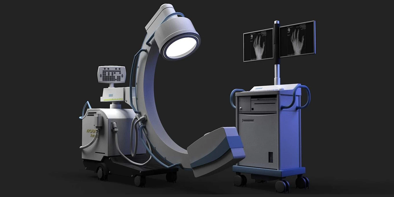
A C-arm machine is an X-ray-based system with both radiographic and fluoroscopic capabilities that provide users with high-quality, minimally invasive, and real-time images of the body. Such capabilities make the C-arm a useful and versatile companion for surgeons and physicians performing complicated tasks requiring speed and accuracy simultaneously.
Key components of a C-arm
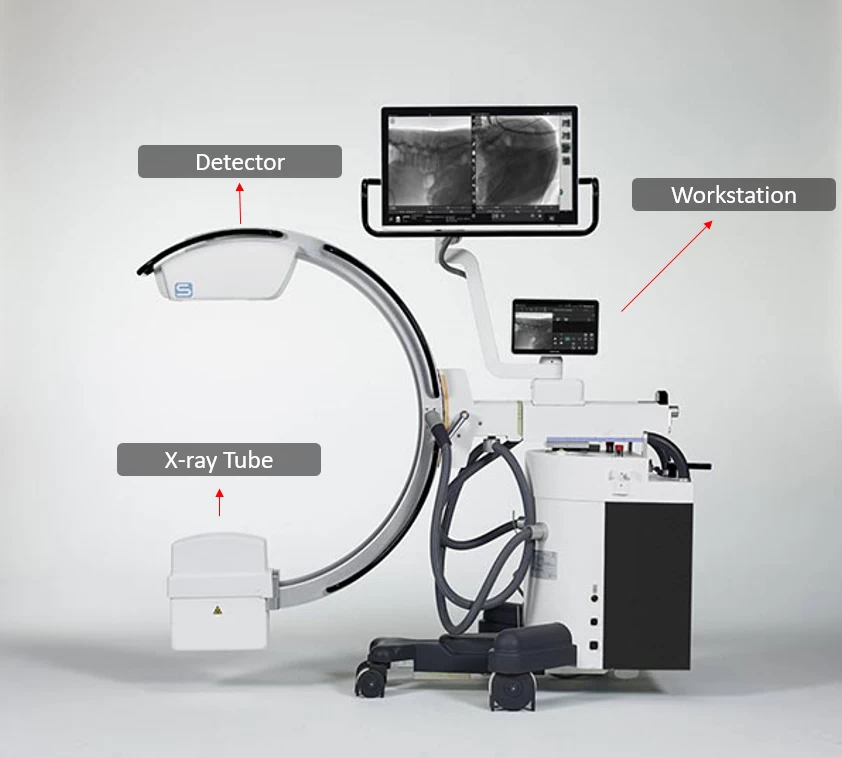
As with any X-ray-based machine, a C-arm is comprised of three key components, an X-ray tube, a detector, and a workstation. The C-shaped arm, where the name “C-arm” is derived from, is the part connecting the tube and the detector. This arm allows horizontal, vertical, and rotational movement so images can be acquired at optimum angles and distances.
X-ray generator
The X-ray generator used in a C-arm is not so different from the ones used in normal radiography units. It performs the task of emitting X-rays, which travel through the patient’s body and reach the detector. The output of the tube can be modified by the operator via the workstation at any given time providing excellent flexibility in image contrast and brightness.
Detector
As the name suggests, a detector detects X-rays and converts them into images. There are multiple types of detectors, but modern X-ray machines, especially C-arms, use flat panels.
Flat-panel detectors are the most common detector type used in digital radiography. They convert X-rays into electrical charges, directly or indirectly, and these charges are then read out and used to create the final image. Compared to image intensifiers used in older models, flat panels are lighter, smaller, more durable, and less susceptible to artifacts and distortions.
Workstation
While an X-ray generator and detector are the beating heart of a C-arm, the workstation is the component that has the most interaction with the user. It must be user-friendly while providing a wide range of functions. The workstation contains several handles controlling the movement and positioning, multiple switches and buttons handling exposure settings, one or more monitors, advanced software for image visualization and manipulation, etc.
Mobile C-arm
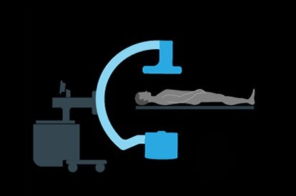
When trauma occurs or surgery is in progress, it is vital that the patient’s movement is at its minimum. This is when a mobile C-arm shines.
A mobile C-arm is composed of the same components as a normal, static C-arm. It provides fluoroscopic and radiographic modes, but the main difference is its ability to move. This movement allows convenient positioning of the machine while keeping the patient still. It also introduces new hardware into the operation room and adds the line of sight constraints imposed by optical tracking systems. The availability of a mobile C-arm can have a great positive impact on the accuracy and efficiency of different procedures, especially high-precision operations.

Types of C-arm
Different applications require different sizes of machinery. To answer the needs of different situations, C-arms are produced in a variety of sizes including Mini, Compact, Full-size, and Super C-arms.
Mini C-arm
A mini C-arm is smaller in size and provides great flexibility for performing studies of extremities like feet, knees, hands, and elbows.
Compact C-arm
Bigger than a mini C-arm, and smaller than a full-size C-arm, a Compact C-arm is capable of imaging larger organs while preserving space and mobility.
Full-size C-arm
Full-size C-arms are the most popular due to their ability to provide images of any organ. A full-size C-arm can have an open space of 26 inches, enabling the user to fit a table and patient between tube and detector.
Super C C-arm
While they need a spacious room, their biggest merit is the 33 inches of open space, making them suitable for larger or obese patients, as well as operations that need more usable areas to operate and image at the same time.
3D Mobile C-arms
Traditional C-arms typically provide radiography and fluoroscopy modes, leading to 2D images of the object. As the need for higher accuracies became prevalent, 2D images failed to satisfy such need. This is where 3D C-arms were invented. A mobile 3D C-arm is an upgraded version of customary C-arms. Not only, It can provide axial, coronal, and sagittal images, but also it is capable of producing 3D reconstructions of the object. These 3D visualizations can better demonstrate the relation between a lesion and its surrounding tissue, the placement accuracy of implants, etc.


Applications
C-arm is well-integrated into healthcare, providing a wide range of applications, ranging from orthopedic surgeries to pain management.
Orthopedics
C-arm has revolutionized the field of orthopedics as it provides quick access to high-quality and detailed images on the spot. Outcomes of orthopedic surgeries for alignment of fractures, placement of implants, joint replacements, etc, have benefited from exceptional properties of C-arm in terms of increased accuracy and efficiency.

Fluoroscopy
Real-time visualization of internal organs and structures is vital for tasks of angiography, urology, gastroenterology, and other complicated surgeries like stent insertions and catheter placements. C-arm machines with fluoroscopy modes can provide these online images and help increase the success rate while reducing procedure time, as longer times can increase the chance of side complications. This is of more importance in emergency medicine.

Pain management
When targeted treatment is needed, C-arm brings high-resolution and real-time images to the patient’s bedside. This allows physicians to perform guided injections such as inter-articular injections with higher precision and more convenience, bringing more effective relief from acute or chronic pains.

C-arm has become an inseparable part of many surgeries. It makes high-resolution radiography and fluoroscopy more accessible, improving the accuracy and confidence of different procedures while helping doctors save valuable time.