پرتوهای ایکس نوعی از امواج الکترومغناطیسی هستند که قابلیت نفوذ به درون اجسام را دارند. اینکه پرتو چه میزان در جسم نفوذ کند، از آن عبور کند یا نه، چه میزان عبور کند، جذب شود یا نه و چه میزان جذب شود، بستگی به ویژگیهای ماده از جمله عدد اتمی و چگالی آن دارد. بنابراین میزان جذب پرتو در مواد مختلف متفاوت است. مواد با عدد اتمی کوچکتر و چگالی کمتر، جذب کمتری دارند. از پرتوی ایکس در پزشکی برای تصویربرداری از استخوانها و برخی اندامها استفاده میشود. این پرتوها همچنین در درمان برخی تومورها کاربرد دارند.

برای انجام تصویربرداری، بیمار بین یک منبع پرتوی ایکس و یک آشکارساز پرتو قرار میگیرد. پرتو از منبع به سمت بیمار تابیده میشود و پس از عبور از آن به آشکارساز میرسد. بسته به نوع بافتی که در مسیر پرتو قرار دارد، میزان مخشصی پرتو به آشکارساز میرسد. بنابراین نقشه ای از میزان تضعیف پرتو توسط بافت، روی آشکارساز به صورت سیاه و سفید و خاکستری تشکیل میشود. برای مثال استخوان که به علت وجود کلسیم در ترکیباتش که عدد اتمی نسبتا بالایی دارد، چگالی بالا و جذب بالاتری دارد، در تصویر (اغلب) روشنتر و نزدیک به سفید ظاهر میشود. اگر استخوان دارای ترک و شکستگی باشد (وجود هوا و فضای خالی)، این شکستگی به رنگ سیاه و تیره در تصویر روشن استخوان، به وضوح دیده میشود. ریهها نیز به علت وجود هوا و حفرات به صورت تیره دیده میشوند، اما اگر عفونت و یا توموری در ریهها وجود داشته باشد چون چگالتر از هوا هستند به صورت لکههای روشن روی تصویر ریه دیده میشوند.

برخی کاربردهای پرتوی ایکس در تشخیص پزشکی
- تشخیص شکستگیها
- بررسی عفونتها
- بررسی کلسیفیکیشن (برای مثال سنگ کلیه)
- بررسی برخی تومورها
- بررسی آرتروز مفاصل
- تشخیص پوکی استخوان
- کاربردهای دندانپزشکی
- تشخیص انسداد عروق
- تشخیص وجود اجسام خارجی در بدن (بلعیدن اجسام کوچک توسط خردسالان)
- و …
انواع تصویربرداری پزشکی با استفاده از پرتوی ایکس
رادیوگرافی معمولی: شکستگی استخوان، تومورهای خاص و سایر تودههای غیر طبیعی، ذات الریه، برخی از انواع جراحات، کلسیفیکاسیونها، اجسام خارجی، مشکلات دندانی و غیره را تشخیص میدهد. این روش تمام بافتهای موجود در مسیر را در یک صفحهی دو بعدی تصویر میکند، بنابراین امکان بررسی اجزای داخلی با دقت کمتری امکانپذیر است. برای رفع این مشکل روش سی تی ابداع شد.
سی تی اسکن یا توموگرافی کامپیوتری: ترکیب رادیوگرافی معمولی با پردازشهای کامپیوتری که مجموعه تصاویر مقطعی از بدن تولید میکند. این تصاویر میتوانند با هم ترکیب شوند و یک تصویر سه بعدی ایجاد کنند. تصاویر سی تی جزئیات بیشتری از رادیوگرافیهای ساده دارند و به پزشکان این امکان را میدهند که ساختارهای داخلی بدن را از زوایای مختلف بررسی کنند.
ماموگرافی: رادیوگرافی از پستان که برای تشخیص سرطان استفاده میشود. تومورها معمولاً به صورت تودههای منظم یا نامنظم ظاهر میشوند که اغلب تا حدودی روشنتر از پسزمینهي رادیوگراف هستند. ماموگرافی همچنین میتواند تکه های ریز کلسیم به نام میکروکلسیفیکاسیون را تشخیص دهد که به صورت لکه های بسیار روشن در ماموگرافی ظاهر می شود. اگرچه میکروکلسیفیکاسیونها معمولاً خوش خیم هستند، اما گاهی اوقات ممکن است وجود نوع خاصی از سرطان را نشان دهند.
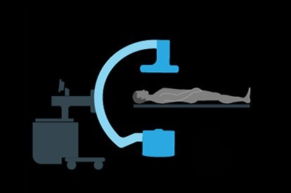
فلوئوروسکوپی: در این روش از پرتوی ایکس و یک صفحه فلوئورسنت برای گرفتن تصاویر لحظهای (زنده) از حرکات داخل بدن یا مشاهدهی فرآیندهای تشخیصی مانند پیگیری مسیر ماده حاجب تزریق شده یا بلعیده شده استفاده میشود. به عنوان مثال: مشاهدهی حرکت قلب تپنده، یا با کمک عوامل کنتراستزا، برای مشاهدهی جریان خون. این فناوری همچنین با کمک یک ماده کنتراستزا برای هدایت یک کاتتر طی آنژیوپلاستی قلب و همینطور در جراحیهای دقیق کاشت ابزار داخل بدن استفاده میشود.
کاربرد پرتوی ایکس در درمان پزشکی
پرتودرمانی در معالجهی سرطان: پرتوی ایکس و سایر انواع پرتوهای پرانرژی را میتوان برای از بین بردن تومورها و سلولهای سرطانی با آسیب رساندن به DNA آنها استفاده کرد. دوز تابشی مورد استفاده برای درمان سرطان بسیار بالاتر از دوز مورد استفاده در تصویربرداری تشخیصی است.


